
Respiratory System
Functions of the Respiratory System
1.Oxygen supplier
2.Elimination
3. Gas exchange
4.Passageway
5. Humidifier
•Nostrils:
- allow air to enter the nasal cavities so it can be purified and
sent to the other parts of the respiratory tract.
•Nasal cavity :
- Forms the interior of the nose. It is the entry point for inhaled
air and the first of a series of structures which form the
respiratory system. The cavity is entirely lined by the nasal
mucosa, which form the physical barriers of the body’s immune
system. These barriers provide mechanical protection from the
invasion of infectious and allergenic pathogens.
•Olfactory receptors:
- located in the mucosa of the nasal cavity. They detect air-borne
odour molecules that enter the nasal cavity
•Respiratory mucosa:
- consists of various types of epithelial cells ranging from ciliated
columnar to simple squamous.
•Mucus:
- produced by the mucosa’s glands that moistens the air and
traps incoming bacteria and other foreign debris
•Nasal concha:
- also called Turbinate, or Turbinal, bony elements forming the
upper chambers of the nasal cavities. They increase the surface
area of these cavities, thus providing for rapid warming and
humidification of air as it passes to the lungs.
•Palate, in vertebrate anatomy, it is the roof of
the mouth separating the oral and nasal cavities
*The hard palate is a bony subsection of the skull that makes up
almost two-thirds of the entire palate. It forms a division
between the mouth and the nasal passages.
The hard palate creates a vacuum inside the mouth to enable
liquid ingestion. Together with the tongue, the hard palate
produces distinctive phonetic sounds, including palatal
consonants such as /ɟ/ and /j/.
*The soft palate is a muscular structure located in the posterior
part of the palate. It is mainly responsible for creating an
incomplete partition between the oropharynx and the mouth.
The soft palate prevents nasal reflux by closing the nasopharynx
during swallowing. Without the soft palate, liquid and solid food
can enter the nasal cavity. Additionally, the soft palate works
with the tongue to produce velar consonants such as [ŋ], [k] and
[g].
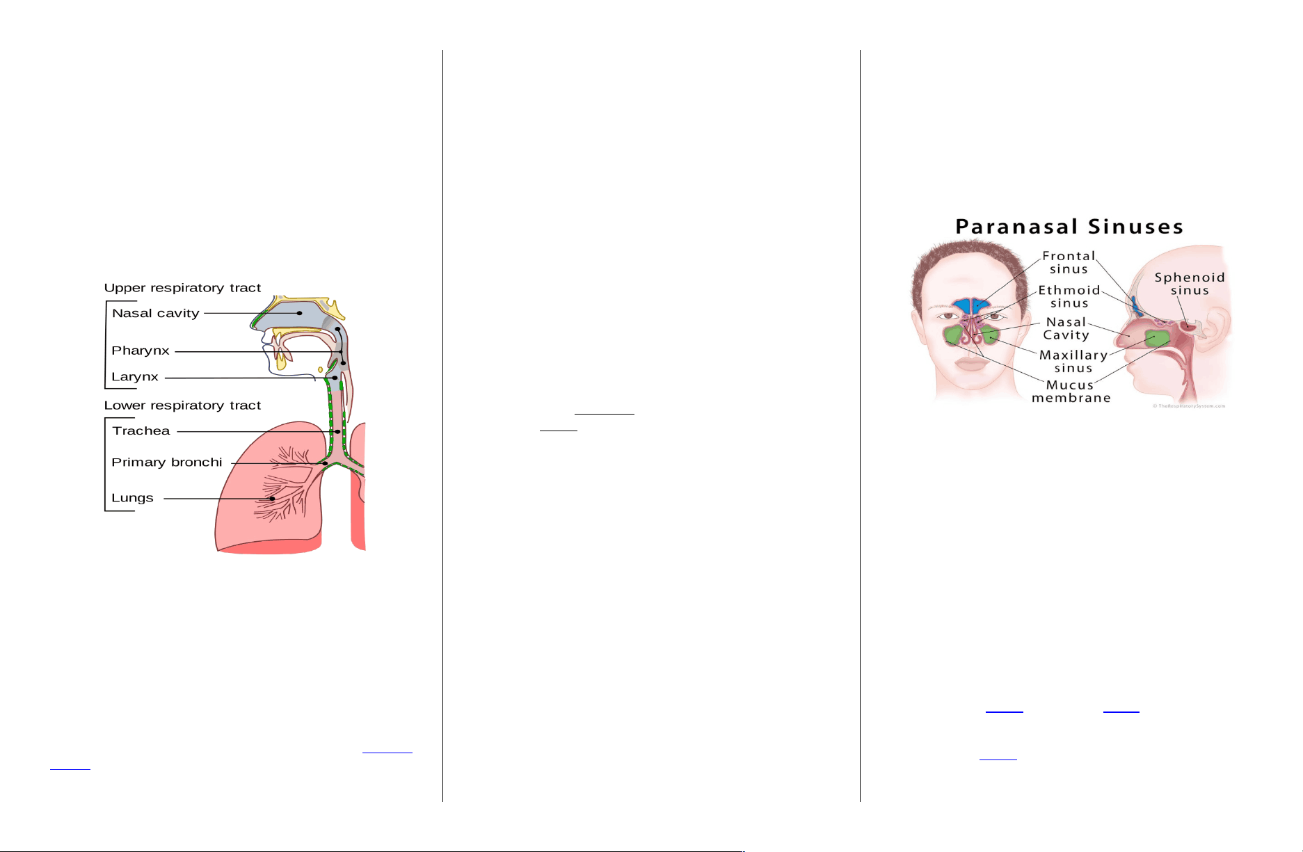
•The paranasal sinuses are air-filled extensions of the
respiratory part of the nasal cavity. There
are four paired sinuses, named according to the bone in
which they are located; maxillary, frontal, sphenoid and
ethmoid.
•The function of the sinuses is thought of humidifying
the inspired air. They also reduce the weight of the
skull.
Pharynx
Size. The pharynx is a muscular passageway about 13
cm (5 inches) long
Function. Commonly called the throat, the pharynx
serves as a common passageway for food and air.
Portions of the pharynx. Air enters the superior
portion, the nasopharynx, from the nasal cavity and
then descends through
the oropharynx and laryngopharynx to enter the larynx
below.
Eustachian tube (pharyngotympanic tube) connects the
middle ear cavity with the nasopharynx..
Palatine tonsils. The palatine tonsils are in the
oropharynx at the end of the soft palate.
Lingual tonsils. lie at the base of the tongue.