FINAL EXAM
REVIEWER IN
ANATOMY AND
PHYSIOLOGY
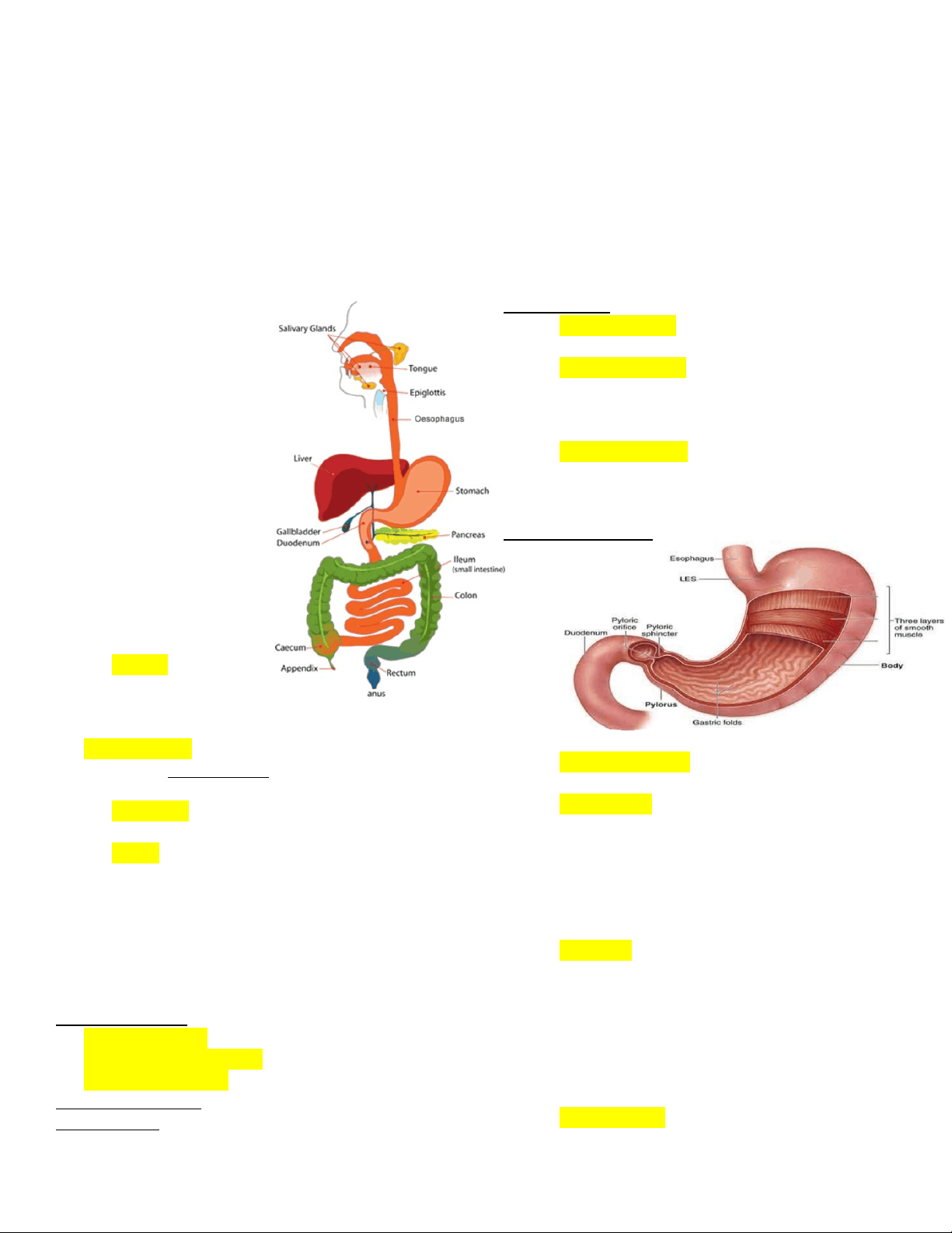
DIGESTIVE SYSTEM
DIGESTIVE SYSTEM FUNCTIONS:
1. Food ingestion
2. Food digestion
3. Nutrient absorption
4. Nutrient metabolism
5. Waste elimination
2 PARTS OF THE DIGESTIVE SYSTEM
1. Gastrointestinal Tract (GI)
2. Accessory Gland
GI/ DIGESTIVE TRACT
1. Oral cavity
2. Pharynx
3. Esophagus
4. Stomach
5. Small intestine
6. Large intestine
GI TRACT HISTOLOGY
1. Mucosa “Inner”
Mucous Epithelium
Lamina Propria
Muscularis Mucosae
2. Submucosa
SUBMUCOSAL GLAND – regulates the
mucus.
3. Muscularis (Smooth Muscle)
Peristaltic/Peristalsis Movement
4. Serosa “outer most layer”
Made up of connective tissue layers
ORAL CAVITY
Parts:
Lips & Cheeks – mastication and speech
Tongue (7th Cranial Nerve)– speech,
taste, mastication, & swallowing
SALIVARY GLANDS
1. Parotid Gland – largest (angel of the jaw)
2. Submandibular Gland
3. Sublingual Gland – behind the tongue
SALIVARIC AMYLASE – enzyme of the saliva
STENSES DUCT – it is where the parotid excretes.
PHARYNX
1. Nasopharynx
2. Oropharynx
3. Hypopharynx
ESOPHAGUS
Connects pharynx to the stomach
Upper esophageal sphincter
Lower esophageal sphincter (cardiac
sphincter/ gastroesophageal sphincter)
– it controls the opening either
proximal or distal.
SWALLOWING:
1. Voluntary Phase – tongue moves bolus of food
from oral cavity to pharynx.
2. Pharyngeal Phase – soft palate close to
nasopharynx & epiglottis closes larynx;
pharyngeal muscles elevate the pharynx and
larynx, and move bolus into the esophagus
3. Esophageal Phase – peristalsis move food to
the stomach.
STOMACH
3 Wall Muscle Layers
Longitudinal
Circular
Oblique
GASTRIC GLANDS
1. Mucous Neck Cells – mucus (protects the
stomach lining)
2. Parietal Cells – Hydrochloric Acid & Intrinsic
Factor.
Vitamin b12 – comes from animal
sources (Ex. Fish, Egg, Milk). Vitamin
b12 helps in red blood cell maturation.
Lack of vitamin b12 can lead to anemic,
etc.
3. Chief Cells – could be found in submucosa
PEPSINOGEN regulates PEPSIN
(enzyme) – it is a strong breaker of
peptide bond.
LACTEAL – absorbs lipids.
REGULATION OF STOMACH SECRETIONS:
- Peripheral Nervous System, Gastrin and
Histamine helps in the increase of secretions.
1. Cephalic Phase – sight, smell, taste & though of
food.